The 2020 Spring Schoenborn Symposium was an Outstanding Success!
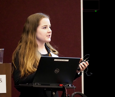
The Schoenborn Symposium is a showcase for the talent and accomplishments of our graduate students.
 The Symposium has two sections. During the first section, senior-level Ph.D. candidates make formal presentations about their research and the associated results. During the second section, mid-level Ph.D. students make informal poster presentations about their research. Faculty judges select first-, second-, and third-place winners of the formal presentations and graduate students select the poster presentation winners.
The Symposium program also includes presentations of the Vivian T. Stannett Fellow Award and the Praxair Exceptional Teaching Assistant Awards.
The 2020 spring winners are:
Formal Presentations
| First Place – Robert Epps (Abolhasani lab) Background: In the development of next-generation photovoltaics and light-emitting diodes, colloidal inorganic perovskite quantum dots (PQDs) have drawn notable attention for their highly tunable bandgap properties, high-charge carrier mobility and defect tolerance, and adaptability towards solution phase processing. However, studies of this material group and other colloidal semiconductor nanocrystals requires extensive exploration of their massive reaction parameter space within highly controlled reaction environments. Conventional flask-based, trial-and-error approaches are, therefore, highly unlikely to effectively capture the full potential and optimal synthesis conditions of these high-priority materials. Further complicating this process, across the accessible bandgap range, optimal synthesis parameters will vary significantly. Flow synthesis platforms have recently been demonstrated as a time- and material-efficient reaction monitoring strategy for synthesis, screening, and optimization of colloidal nanomaterials. The high sampling rate, low chemical consumption, and precise process control (automation) of flow reactors greatly reduces the challenges in exploring complex reaction spaces; however, high-throughput reaction screening technologies alone are likely not able to make significant breakthroughs, due to the massive scope of relevant colloidal synthesis conditions. Results: In this work, we present a modular microfluidic platform integrated with a machine learning (ML)-enhanced reaction optimization algorithm for on-demand synthesis of high-quality inorganic perovskite QDs with desired optical properties. The intelligent QD synthesis platform consists of multiple computer-controlled pumps for on-demand reagent delivery/dosing, and an in-line flow cell for automatic UV-Vis absorption and photoluminescence spectroscopy. With this strategy, we have autonomously optimized reactant compositions for the simultaneous improvement of photoluminescence quantum yield (PLQY) and emission full-width at half-maximum (FWHM) at any desired peak emission energy (PE) – i.e. emission color. These optimizations are performed without any prior experimental information, and they reach desirable PLQY, FWHM, and PE values substantially faster than existing black box optimization techniques – in less than 25 samples. We then apply this same technique but instead pre-train the neural network models with prior experimental data before conducting the adaptive sampling procedure. As a result, we have further pushed the optimal measured values for nine of eleven target PE set points, in spite of performing the synthesis with a completely separate batch of precursors. Conclusions: These techniques not only enable unsupervised material optimization and discovery, but they offer more consistent manufacturing of highly sensitive reactions. Further application of this strategy will advance the quality of commercial nanocrystals and expedite reaction studies, thereby leaving more time and resources available for creative material synthesis exploration. |
|
| Second Place – Ria D. Corder (Khan lab) Background: Biological tissues are complex composite materials whose mechanical properties are often difficult to measure by traditional techniques. Quantification of bulk tissue properties, such as modulus and viscoelasticity, can be used to aid in disease diagnosis and design of novel therapies. We demonstrate the ability to measure multiple types of mammalian tissues, with elastic moduli ranging from 100-10,000 Pa, by dynamic oscillatory rheology on a commercially-available rheometer. We then present results from two case studies involving tumor digestion by injected collagenase enzymes and illustrate how rheology can be used to quantify treatment efficacy. In the first, we quantified the degree of in-vivo digestion of human uterine fibroid tissue (benign tumors) by a single injection of collagenase Clostridium histolyticum. In the second, we measured digestion of xenograft human breast cancer (malignant) tumors grown in mice by repeat injections of liberase (blend of collagenase isoforms.) In both studies, we co-injected Liquogel (LQG) [1], a thermoresponsive polymer that transitions upon heating from an injectable solution to a gel, to reduce diffusion of collagenase from the injection site. Results: All tissues exhibited gel-like rheological behavior. We calculated average tissue moduli and viscoelasticity (tan d) for all tissues. Data collected from reduction mammoplasty human breast tissues isolated from multiple individuals demonstrates that rheology can quantify tissue variability. Repeated freeze-thaw studies reveal the effect of sample history and highlight the need for consistent protocols for handling biological samples. We observed that injections of LQG & collagenase significantly reduced the modulus and increased the viscoelasticity of uterine fibroids compared to both buffer controls and free collagenase injections. The impact of collagenase injections on breast cancer tumors was less intuitive; in some instances, co-injection of LQG & collagenase led to the formation of smaller and stiffer tumors. We hypothesize this observation is due to collagenase softening the local microenvironment and discouraging outward tumor growth. Conclusions: Dynamic rheology is a useful tool for characterizing the bulk modulus of biological tissues and for quantifying the effects of tumor digestion by collagenases. In-vivo tumor imaging and histological staining of parallel tissue samples complement rheological measurements to provide a more complete understanding of treatment effects on tumors. References:
|
|
Third Place – Dilara Sen (Keung lab)Human cerebral organoids reveal early spatiotemporal dynamics and pharmacological responses of UBE3ABackground: Human neurodevelopment and its associated diseases are complex and challenging to study. In particular, prenatal neurodevelopmental periods are hard to access experimentally, especially in humans. The role of UBE3A in Angelman Syndrome and Autism Spectrum Disorder is an archetype for these challenges [1,2]. It is paternally imprinted in parts of the mouse brain, exhibits complex subcellular localization, and is suspected to dynamically establish these patterns in gestation [3,4]. Results: In this work, human cerebral organoids reveal that important spatiotemporal dynamics of UBE3A occur very early in human neurodevelopment. In particular, UBE3A localizes to the nucleus of neurons within a few weeks of organoid culture, with a stark transition established between EOMES and TBR1 cortical layers; these localization patterns are disrupted, and in some cases reversed, in Angelman Syndrome hiPSC-derived organoids. Organoids also exhibit early imprinting of paternal UBE3A within 6 weeks of culture, with topoisomerase inhibitors partially rescuing UBE3A levels in Angelman Syndrome organoids. One inhibitor also exhibits over two weeks of persistent rescue after just a single treatment. Conclusions: This work establishes human cerebral organoids as an important model for studying UBE3A biology and motivates their broader use in understanding complex neurodevelopmental disorders. References:
|
Poster Presentations
| First Place – Kevin Lin (Keung lab)
Background: Technological leaps are often driven by key innovations that transform the underlying architectures of systems. Current DNA-based information storage systems largely rely on polymerase chain reaction, which broadly informs how information is encoded, databases are organized, and files are accessed. Here we show that a hybrid ‘toehold’ DNA structure can unlock a fundamentally different, dynamic DNA-based information storage system architecture with broad advantages. This innovation increases theoretical storage densities and capacities by eliminating non-specific DNA-DNA interactions common in PCR and increasing the encodable sequence space. It also provides a physical handle with which to implement a range of in-storage file operations. Finally, it reads files non-destructively by harnessing the natural role of transcription in accessing information from DNA. This simple but powerful toehold structure lays the foundation for an information storage architecture with versatile capabilities. Results: This set of challenges could be overcome by using a hybrid file structure that included a single-stranded DNA (ssDNA) ‘toeholds’ and double-stranded (dsDNA) payload, and by the way organisms naturally access information from their genome through transcription. With the help of the toehold, we demonstrate that DNA strands that contain information of interest can be separated at room temperature (25 °C), accessed non-destructively through natural transcription driven by a modified T7 promoter sequence, and returned to the file database for recovery using functionalized magnetic beads 4 . In addition, since the accesses are without melting the file templates, non-specific amplification and off-target products are eliminated. This promotes the optimization of encodings, thereby improving oligo availability and information density of the system by orders of magnitude. Furthermore, we demonstrate that the toeholds of specific file strands can bind other appropriate designed oligos, thus providing in-storage file manipulation including file locking, renaming and deletion. Conclusions: The DNA-based information storage system has captured enormous attention recently. Using the hybrid file structure has been shown to increase the theoretical information density and capacity of DNA storage by inhibiting non-specific binding within data payloads, enable transcription-based, non-destructive information access, and make possible in-storage file operations. This work demonstrates dynamic information access and manipulations are practical for DNA-based information storage systems. References:
|
|
| Second Place – Aditya Sapre (Velev lab) Background: As nanomaterials become more common, available, and better understood, they are investigated in various applications, including plant protection. Several research groups have studied the use of different nanomaterials, including metal oxide nanoparticles and carbon nanotubes, as antifungals, but these materials remain in the environment for a very long time with potentially toxic effects. Our group has developed environmentally benign nanoparticles (EbNPs) of lignin, and we are investigating their use as an antifungal agent. Results: Our EbNPs have the potential for a wide range of applications, and have been proven to be efficient antibacterial agents when loaded with silver ions. Preliminary experiments showed that EbNPs were almost as efficient at preventing fungal growth as a commercial chemical, and we are studying the fundamental interactions between all the components to understand these results. Through the use of bright-field and fluorescence microscopy, we are able to track the trends of fungal growth and where the EbNPs are going on the spores or substrate. Using various combinations and concentrations of EbNPs, spores, and different types of surfactants, we investigate colloidal interactions in solution, coffee ring formation on surfaces, and infectivity when applied to rose petals. We are also studying the spores and nanoparticle interactions over agar plates to macroscopically observe the difference between the systems. Conclusions: EbNPs are interacting with fungal spores causing aggregation which is resisting the spore development. Further studies are currently ongoing to better understand the phenomenon. Such essential understanding of these interactions will assist in formulating efficient and environmentally benign antifungal products. References:
|
|
| Third Place – Rajesh Paul (Wei lab) Background: Molecular diagnoses of plant pathogens play a vital role for global crop protection, and the first step of molecular diagnoses is to isolate DNA from infected plant specimens. However, DNA isolation from plant cells is a complicated and time-consuming process due to the presence of rigid polysaccharide cell walls. As a result, isolation of high-quality plant DNA is mainly confined to well-equipped laboratories, and sample preparation becomes one of the major hurdles to perform molecular diagnostic assays of plant pathogens in the field. To expedite sample preparation and pathogen detection on site, a simple and rapid DNA extraction method has been developed using a polymer microneedle (MN) patch to isolate genomic or pathogenic DNA from plant leaves. Results: MN patch-based DNA extraction method is minimally invasive, and isolates polymerase chain reaction (PCR) amplifiable DNA within one minute from several plant species such as tomato, potato, pepper, and banana. MN patches used for DNA extraction, are made of polyvinyl alcohol (PVA), and each patch consists of 225 microneedles. These conical microneedles penetrate deep into plant tissue and break hard-to-lyse plant cell walls to release encapsulated nucleic acid materials. For DNA extraction, a MN patch is gently pressed on the surface of the leaf for few seconds, and then, the patch is peeled off and rinsed with 100 µL TE buffer to collect absorbed DNA from microneedles. MN patches successfully extract Phytophthora infestans DNA from both laboratory-inoculated and field samples for detection of late blight disease in tomato. For laboratory-inoculated samples, the MN patch-based DNA extraction method achieved 100% detection rate compared to cetyltrimethylammonium (CTAB)-based DNA extraction method when the samples were taken 3 days after inoculation. For field samples, the MN extraction method successfully detected P. infestans in all blind samples tested from older lesions. Conclusions: MN patch-based DNA extraction method reduces sample preparation time from 3-4 hours in a conventional assay to ~1 minute, and extracted DNA is directly applicable for amplification reactions without further purification. For applications, we have demonstrated that MN extraction detects P. infestans DNA in both inoculated and field-collected tomato leaf samples. Moreover, this simple and rapid extraction method could be used to isolate other plant pathogens’ DNA from various plant species directly in the field, and thus have great potential to become a universal sample preparation technique for on-site molecular diagnosis of plant pathogens. References:
|
|
View the complete Symposium program here
Dr. Caryn Heldt (Ph.D., 2008) delivered the Keynote talk, “How I got excited about viruses and their surface interactions.” Dr. Heldt is the Director of the Health Research Institute, the James and Lorna Mack Chair in Bioengineering, and an Associate Professor of Chemical Engineering at Michigan Technological University.
The Vivian T. Stannett Fellow Award is also presented at the Symposium. It’s named in memory of Professor Vivian T. Stannett, a CBE faculty member who was an internationally renowned polymer scientist, research leader, and member of the National Academy of Engineering. The Award recognizes research excellence, initiative, focus and tenacity during the early careers of Ph.D. candidates in the department. The 2020 Fellow Award winner is Kyle Tomek. Rajesh Paul and Jiaqi Yan were Honorable Mention recipients.
The Fall 2020 Praxair Exceptional Teaching Assistant Award was presented to Rachel Nye. Joseph McCaig is the Spring 2019 Praxair Award winner. The Award recognizes instructional effectiveness and management skills of Ph.D. candidates who serve as exemplary teaching assistants in CHE courses. The Award recipient goes above and beyond the call of duty and provides students with tireless and selfless attention to high-quality instruction and professionalism.
Congratulations to the Schoenborn competition participants and winners, and the Stannett and Praxair award winners. Well done to all!
- Categories: