Daniel Kuncicky
Ph. D., Chemical Engineering, 2007 North Carolina State University B. S., Chemical Engineering, 2001University of Florida, Gainesville, Florida |  |
Research Focus: On-line SERS Detection on Templated Gold Substrates
My research is focused on developing a surface enhanced Raman spectroscopy (SERS) based sensor that targets chemical and biological agents. Raman spectroscopy allows for in situ real time measurements, non-destructive and non-intrusive sampling, remote sampling through fiber optics, and detection of analytes in aqueous solvents. In addition, the spectra are well-resolved with high information content. However, poor sensitivity inhibits the detection of low concentrations of harmful chemical and biological agents. This problem can be solved by adsorbing the analyte onto nanostructured gold and silver surfaces. This can lead to Raman spectra that are a million times more intense than ones without surface enhancement. The predominant mechanism of Raman enhancement is widely believed to stem from the electric field enhancement that occurs near small interacting metal particles illuminated with light that excites the surface plasmons in the metal. The fabrication of such substrates is a major challenge in SERS spectroscopy.We have developed a technique for making highly enhancing and stable SERS substrates by using colloidal crystals to template gold nanoparticles into structured porous films. It is expected that the size, shape, and spacing of the metal particles will influence the electric field enhancement. Currently, I am focusing on optimizing the performance of the SERS substrates, and as a first step I am studying how the film structure affects the intensity of the Raman spectra. A new microphotonic method is being developed that will allow us to directly visualize the position, shape and strength of the plasmon resonance.

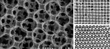
Figure 1: Electron micrographs of SERS films showing the hierarchical structure of individual 20-30 nm Au particles arranged around micron-sized periodically spaced cavities (left) and square bilayer (upper right) and hexagonal close packed (lower right) structures which are examples of 3-D ordered Au structures which can be fabricated using the colloidal templating technique.
The efficacy of the SERS films is probed using a flow chamber that continuously samples a flux of liquid. This experimental set-up has the added advantage of being directly applicable as a SERS sensor in chemical detection for military and civilian purposes. Currently, sodium cyanide is used to characterize the SERS activity of the substrates and future work will focus on phosphororganic agent simulants. We will fabricate and characterize substrates that specifically target chemical and biological agents and incorporate them into microscopic sensors with constant sampling capabilities.

Figure 2: Schematic diagram of the experimental system used to evaluate the performance of SERS substrates. The SERS substrate is integrated withing a microfluidic flow chamber and interfaced to the Raman spectrometer.
Publications
